See It. Treat It. Relieve It.
We use live imaging to guide every step of your treatment.

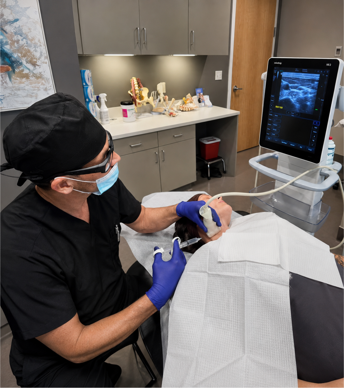
Interventional Orthopedics of Washington uses real-time ultrasound and, when appropriate, fluoroscopy to help the physician visualize the target anatomy and guide the needle to the intended joint, tendon, ligament, spinal structure, or surrounding soft tissue. The injected treatment may include platelet-rich plasma (PRP), bone marrow concentrate, prolotherapy, or another option selected after a clinical evaluation and imaging review.
Image guidance supports precise needle placement, but it does not guarantee pain relief or a specific outcome. A consultation and imaging review are required to determine the appropriate procedure, discuss alternatives and potential risks, review medication instructions, and set realistic expectations. Procedure preparation, the use of contrast, and sedation needs vary by treatment area and medical history.
At Interventional Orthopedics of Washington, all injections are performed using real-time ultrasound or fluoroscopy (live X-ray) to improve accuracy and safety. Image guidance allows our providers to visualize the anatomy being treated and place injections directly at the source of pain rather than relying on surface landmarks.
Diagnostic injections, performed under ultrasound and or fluoroscopic guidance, may be recommended after a new patient consultation to help identify specific pain generators in joints, ligaments, tendons, or the spine. These diagnostic procedures are typically scheduled for a future visit following the initial evaluation.
All injection-based therapies, including platelet-rich plasma (PRP) therapy, cellular (stem cell) therapy, and prolotherapy are always performed under image guidance. Studies show that injections performed without imaging can miss the intended target in up to 30-40% of cases, while image-guided injections exceed 95% accuracy, leading to more reliable outcomes.
Please note: Treatments of any kind, including diagnostic injections, are not included in the new patient initial consultation fee.
Using advanced imaging technology, we deliver precise, minimally invasive injections to the exact source of your pain—enhancing accuracy, safety, and overall treatment effectiveness.
Our real-time imaging allows us to see exactly where we’re treating—helping improve outcomes and minimize risks.
We use live imaging to guide every step of your treatment.
Precision targeting means less trauma, less downtime, and faster recovery.
Every injection is tailored to your unique anatomy and condition.
We use advanced imaging to locate the exact source of your pain.
The needle is guided in real-time to the exact area that needs treatment.
The medication is delivered precisely where it’s needed for maximum effect.
Accurate treatment leads to better pain relief, improved function, and faster recovery.
Clear answers about how image-guided injections work, why precision matters, and how IOW uses ultrasound or fluoroscopy to target the true source of pain.
Image-guided injections are procedures performed with real-time imaging, such as ultrasound or fluoroscopy, to help place treatment precisely into the intended joint, tendon, ligament, nerve space, or other pain-generating structure.
Precision matters because pain often comes from very specific structures. Image guidance allows the physician to see the target area, avoid nearby sensitive tissues, and confirm more accurate placement than a blind injection.
IOW uses ultrasound and fluoroscopic guidance depending on the area being treated and the goal of the procedure. Ultrasound is often used for soft tissues, nerves, and many peripheral joints, while fluoroscopy can be used for deeper structures and spine-related procedures.
They can be both. Diagnostic injections may help confirm which structure is causing pain. Therapeutic injections may deliver regenerative treatments or other targeted solutions to support healing, reduce irritation, or improve function.
Image-guided injections may be used for joint pain, tendon or ligament injuries, arthritis, nerve irritation, spine-related pain, and instability-related conditions. The exact approach depends on your diagnosis, imaging, symptoms, and treatment goals.
The treatment area is cleaned and prepared, then imaging is used to guide needle placement. Most procedures are performed in-office, and your physician will explain what is being targeted before the injection begins.
When performed by trained physicians using appropriate imaging guidance, these injections are designed to improve accuracy and safety. As with any procedure, risks vary depending on the treatment area and injection type, which will be reviewed during your evaluation.
Schedule your discovery call and let’s explore the next best step for your condition.
Medically reviewed by Dr. Otoño Silva, M.D.
Board Certified in Physical Medicine & Rehabilitation · Fellowship-Trained in Interventional Orthopedics, Centeno-Schultz Clinic
Last reviewed